The image shown is a photomicrograph of a transverse section of part of the gas exchange system.
What describes the image?
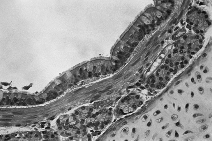
1 )
a thin inner layer of ciliated epithelial cells on top of a layer containing cartilage, supported by elastic fibres
2 )
a very thin epithelial lining with walls containing elastic fibres, surrounded by many blood vessels
an inner layer of ciliated epithelial and goblet cells on top of elastic fibres, supported by an outer layer consisting of cartilage
4 )
an inner layer of ciliated epithelial and goblet cells on top of loose tissue with mucous glands, supported by a continuous ring of cartilage
تحلیل ویدئویی تست
منتظریم اولین نفر تحلیلش کنه!


پرسش و پاسخ های مشابه
سوال کنید یا به سوالات دیگران پاسخ دهید ...